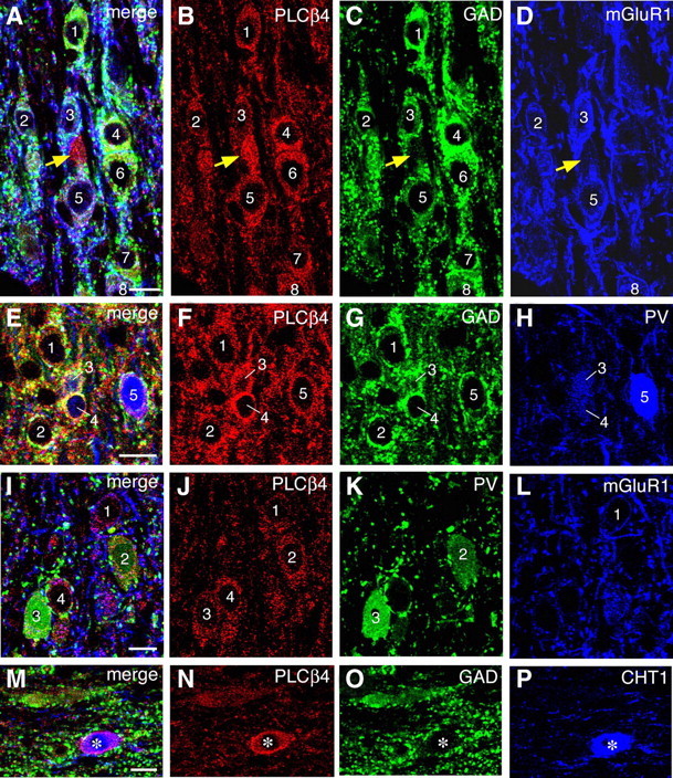
Figure 8.

Triple immunofluorescence showing that PLC-β4 is detected in GABAergic neurons expressing mGluR1a and PV and in cholinergic neurons expressing CHT1. Triple labeling for PLC-β4, GAD, and mGluR1 (A–D), PLC-β4, GAD, and PV (E–H), PLC-β4, PV, and mGluR1 (I–L), and PLC-β4, GAD, and CHT1 (M–P). Labeled neurons are indicated by numerals. Note infrequent and intense PLC-β4 labeling in GAD-negative neuronal elements (A–D, yellow arrows), which are likely to represent cholinergic neurons expressing CHT1 (M–P). Scale bars, 10 μm.
