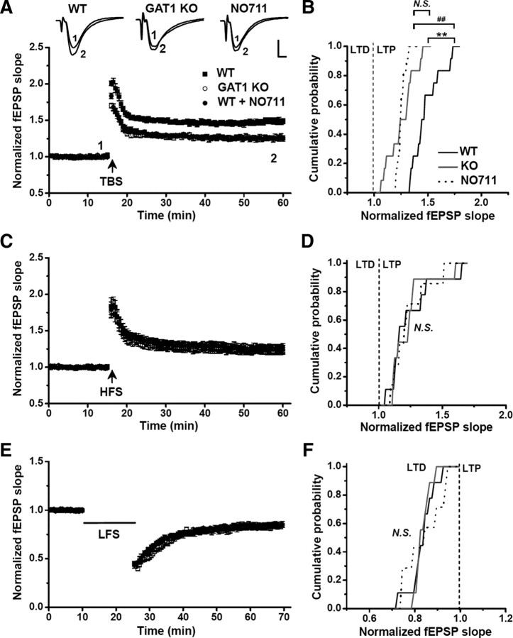
Figure 1.
GAT1 disruption selectively impaired TBS-induced LTP. Field EPSPs were recorded in the CA1 area of hippocampal slices derived from WT and GAT1 KO mice, in the absence of the GABAAR antagonist. The fEPSP slope was normalized to the baseline value before LTP or LTD induction. The magnitude of LTP or LTD was measured as the averaged value from the last 10 recordings. A, Either GAT1 deletion or GAT1 inhibitor NO711 significantly impaired TBS-induced LTP in hippocampal slices (n = 12 for each group). Typical fEPSP recordings were shown before TBS (1) or 40 min after LTP induction (2). B, Cumulative probability of TBS-induced LTP magnitudes for each group. C, D, HFS-induced LTP in slices from GAT1 KO mice, or slices from WT mice in the absence or presence of NO711 (WT, n = 9; KO, n = 9; NO711, n = 7). E, F, LFS-induced LTD in slices from GAT1 KO mice, or slices from WT mice in the absence or presence of NO711 (WT, n = 9; KO, n = 9; NO711, n = 7). For all NO711 groups, 20 μm NO711 was applied throughout the entire experiments. **p < 0.01; ##p < 0.01; N.S., no significant difference; Kolmogorov–Smirnov tests. Calibration: 5 ms, 0.5 mV.

