Figure 4.
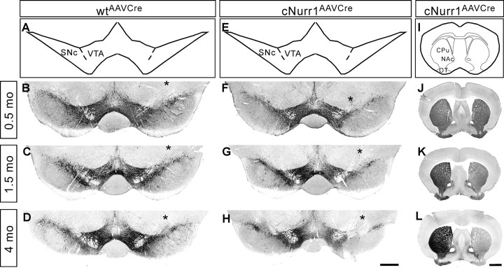
TH expression in both the ventral midbrain and striatum is progressively lost in the injected, but not non-injected, side of cNurr1AAVCre mice. A–H, Sections from 0.5, 1.5, and 4 month (mo; as indicated) old AAV–Cre-injected controls (wtAAVCre) or cNurr1AAVCre mice were used for analyses by nonfluorescent DAB TH immunostaining in the ventral midbrain. The analyzed region within the ventral midbrain is schematically illustrated in A and E. The site of injection, marked by an asterisk in B–D and F–H, was verified in all animals by high-power magnification microscopy and was identified as a small area of injection-induced necrosis. Results show that TH immunostaining is not drastically altered at 0.5 months but is progressively decreased at 1.5 and 4 months in the injected SNc and VTA. I–L, DAB TH staining at the level of striatum. Analyzed regions are indicated in I. TH staining is progressively decreased at 1.5 and 4 months in the side that is ipsilateral to the side of AAV–Cre injection in cNurr1AAVCre mice (J–L). OT, Olfactory tubercle. Scale bars: A–H, 600 μm; I–L, 1 mm.

