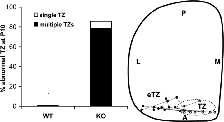
Figure 4.
Abnormally positioned termination zones of temporal RGC axons in ALCAM null mutants. Left, Percentage of WT (n = 10) and ALCAM null mutant (n = 14) mice with abnormally positioned TZs in the SC after DiI injections into the temporal retina at P8 and analysis at P10. Right, Schematic diagram of the location of TZs (open circles) and aberrant (single displaced or multiple) eTZs (filled circles) in ALCAM null mice. The centers of TZs and eTZs from mutant mice (n = 14) are marked and connected. The range of location of TZs of WT axons in the SC is depicted by the dotted oval. L, Lateral; M, medial; A, anterior; P, posterior.

