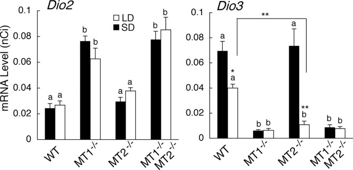
Figure 2.
Effects of targeted disruption of melatonin receptor types on the expression of Dio2 and Dio3 in the ependymal cell layer lining the infundibular recess of the third ventricle. C3H wild-type (WT) and C3H mice with targeted disruption of MT1 (MT1−/−), MT2 (MT2−/−), and both receptor types (MT1−/− MT2−/−) were kept either under short-day (SD, black bars) or under long-day (LD, white bars) conditions for 2 weeks. Different characters (a, b) indicate the significance in each lighting condition (one-way ANOVA followed by Fisher's least significant difference post hoc test, p < 0.05; n = 5–6). Asterisks indicate significant differences between mice kept under SD and LD in each genotype or between mice of different genotypes (t test, *p < 0.05, **p < 0.01; n = 5–6). Error bars indicate SEM.

