
Figure 5.
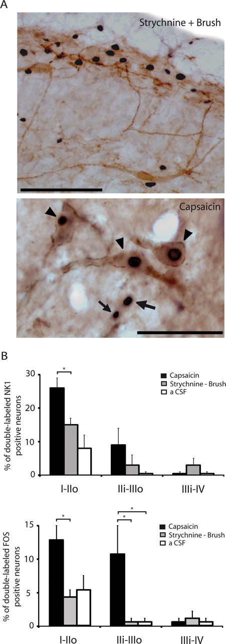
Rare NK1 receptor-expressing dorsal horn neurons are activated by brushing under glycine disinhibition compared with capsaicin injection. A, Images of Fos-positive cell nuclei (black nuclear staining) and NK1 receptor-expressing neurons (brown staining) in lamina I–III of the MDH. Top, After brushing under glycine disinhibition; scale bar, 100 μm; bottom, after capsaicin injection, where clear examples of double-labeled neurons (arrowheads) and Fos-positive cells that are NK1 receptor negative (arrows) are illustrated; scale bar, 20 μm. B, Bar histograms summarizing the percentages of NK1 receptor-expressing neurons and Fos-positive neurons that are double-labeled in the different laminae of the MDH (n = 3–4/group). Fos expression was induced by light brushing of the ipsilateral lip after intracisternal strychnine or by capsaicin injection into the upper ipsilateral lip. Control animals (aCSF) received intracisternal aCSF and brushing without strychnine. I–IIo, Laminae I and outer II; IIi–IIIo, laminae inner II and outer III; IIIi–IV, laminae inner III and IV; *p < 0.05.