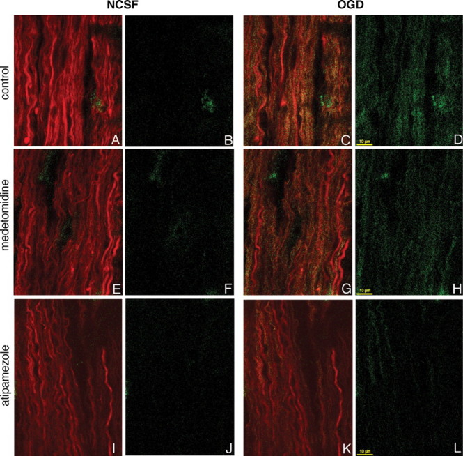
Figure 4.

Confocal images of live rat optic nerve axons coloaded with Alexa Fluor 594 dextran (red) and Ca indicator Fluo-4 dextran (green) in vitro during perfusion in normal CSF (left panels without OGD: A, E, I, both channels; B, F, J, Ca2+-sensitive fluorescence) and after 30 min of exposure to OGD (right panels: C, G, K, both channels; D, H, L, Ca-sensitive fluorescence). Pretreatment with medetomidine (G, H) or atipamezole (K, L) reduced OGD-induced axonal Ca2+ accumulation.
