
Figure 2.
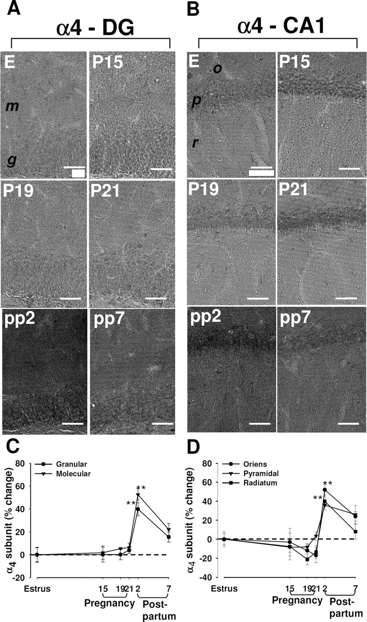
Changes in immunoreactivity for the α4 subunit of the GABAA-R in the rat hippocampus during pregnancy and after delivery. A, B, Representative immunohistochemical images of the distribution of the α4 subunit in the dentate gyrus (A) and in the CA1 region (B) in hippocampal sections from rats in estrus (E), at P15, P19, or P21, or at pp2 or pp7. m, Molecular layer of the dentate gyrus; g, granule layer of the dentate gyrus; o, stratum oriens of the CA1 region; p, pyramidal of the CA1 region; r, radians of the CA1 region. Scale bar, 50 μm. C, D, Images similar to those in A and B were subjected to semiquantitative measurement of α4 subunit immunoreactivity in the granular and molecular cell layers of the dentate gyrus (C), and in the stratum oriens, pyramidal cell layer, and stratum radiatum of CA1 (D). Data are expressed as the percentage change in gray-scale values relative to the corresponding value for rats in estrus (control) and are means ± SEM from six to eight animals at each time point. **p < 0.01 versus estrus (one-way ANOVA followed by Scheffe's test).