
Figure 3.
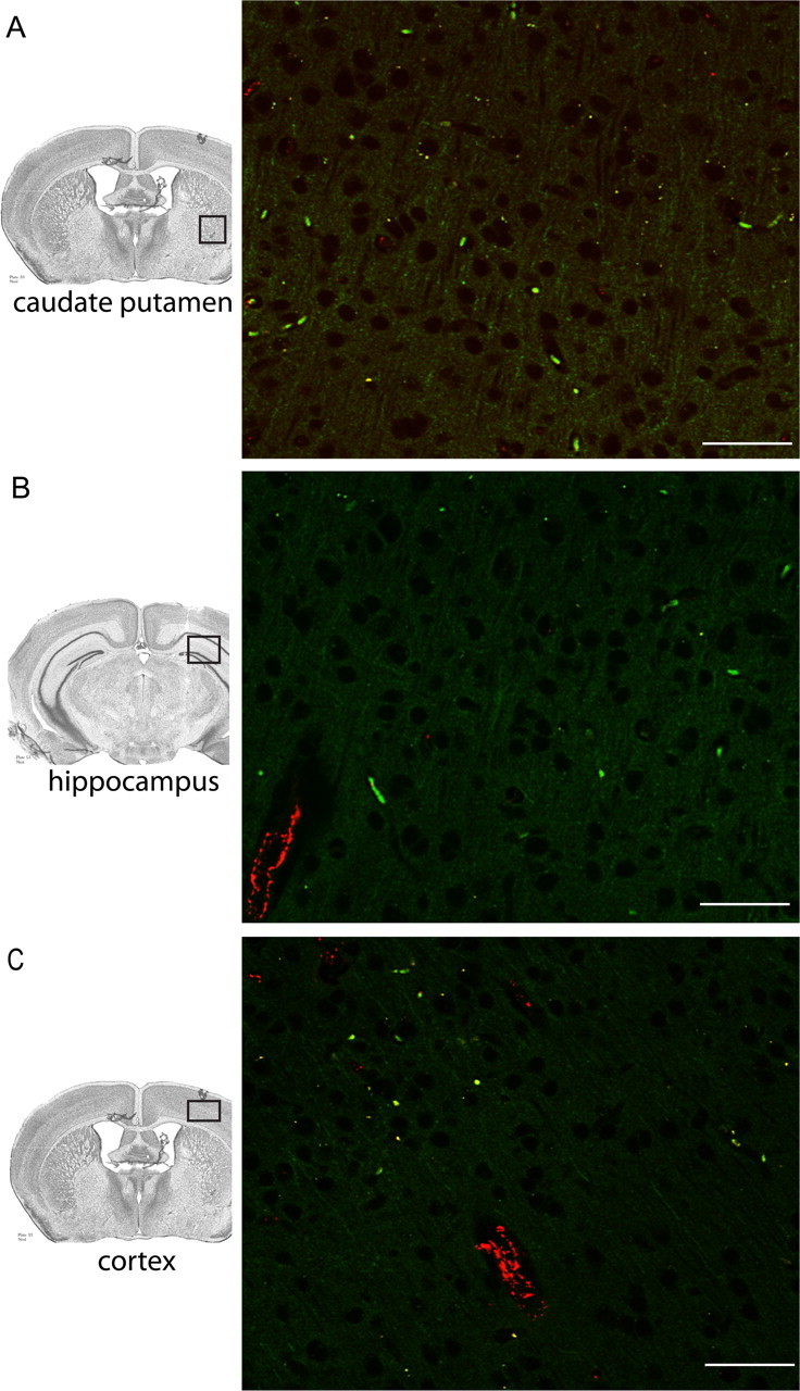
Region-specific detection of CFSE-labeled monocytes in BDR mice. Large numbers of CFSE-labeled monocytes were most readily detected in the cerebral parenchyma of BDR mice in specific regions of the brain, including the basal ganglia (A), hippocampus (B) and motor cortex (C). Immunostaining for vWF was used to identify blood vessels in all three panels. Positive VWF staining is depicted by red immunofluorescence. A, B, C, Brain sections from n = 3 day 10 BDR mice. Scale bar, 20 μm. BV, Blood vessel.