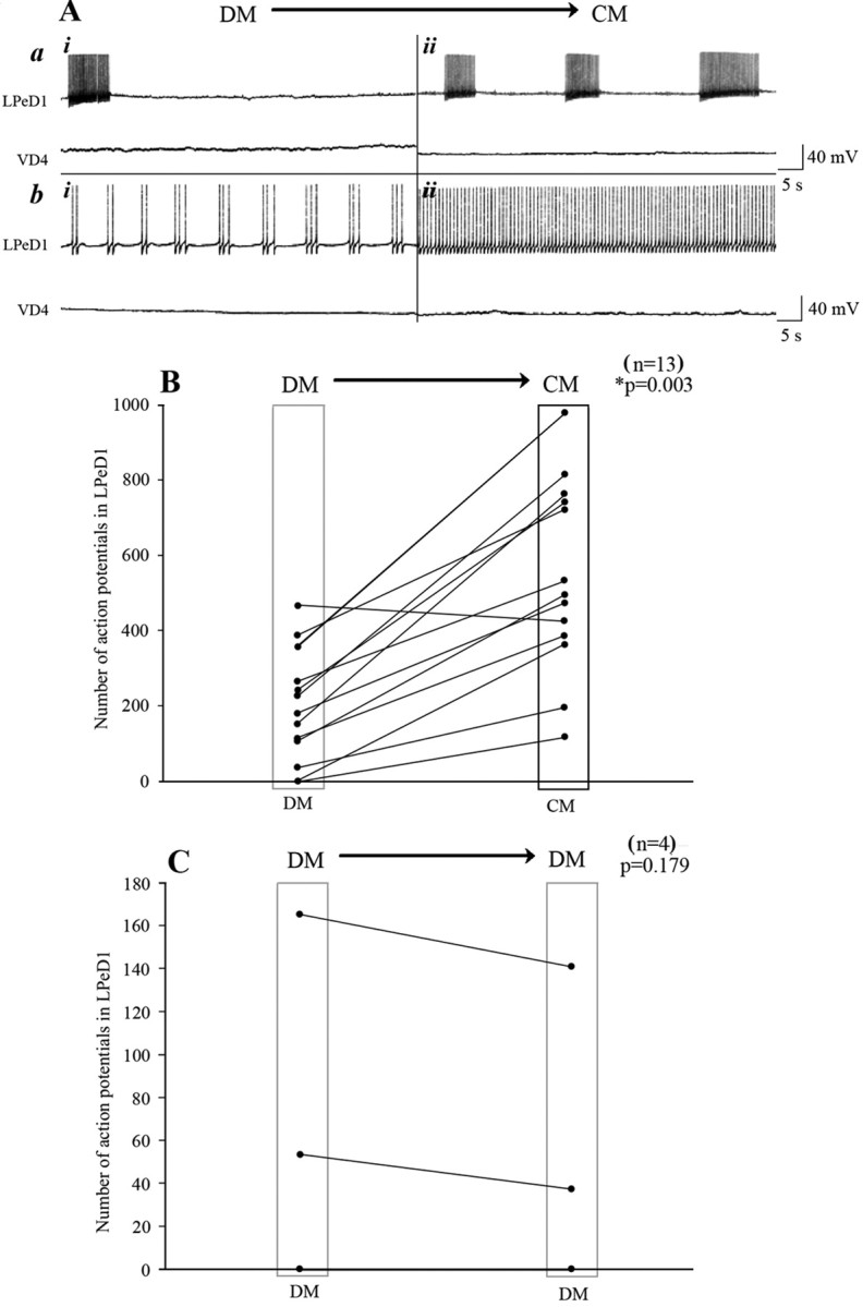
Figure 2.

CM enhanced spontaneous electrical activity of the postsynaptic but not the presynaptic cell. To test whether CM affects the activity patterns of LPeD1 and VD4, cells were first paired overnight in DM, and on day 2, both neurons were impaled with sharp intracellular electrodes. A baseline for spontaneous activity was established for 20–30 min in DM (Ai), and DM was replaced with CM (Aii). Within minutes of CM perfusion, the spontaneous activity of LPeD1 was significantly enhanced (A). Specifically, regardless of LPeD1 's activity status, i.e., bursting (Aa) or tonic activity (Ab), the total number of action potentials increased significantly in CM. In contrast, the CM perfusion did not affect the activity of the presynaptic cell VD4 (A). B, C, Summary data showing the total number of action potentials recorded from LPeD1 [DM to CM (B) and DM to DM (C)]. The spontaneous activity in 93% of LPeD1 cells was significantly enhanced over a recording period of 20 min, whereas DM to DM perfusion did not affect the total number of action potentials in LPeD1 (n = 4). The asterisk indicates significant difference (t test; p < 0.05).
