Figure 4.
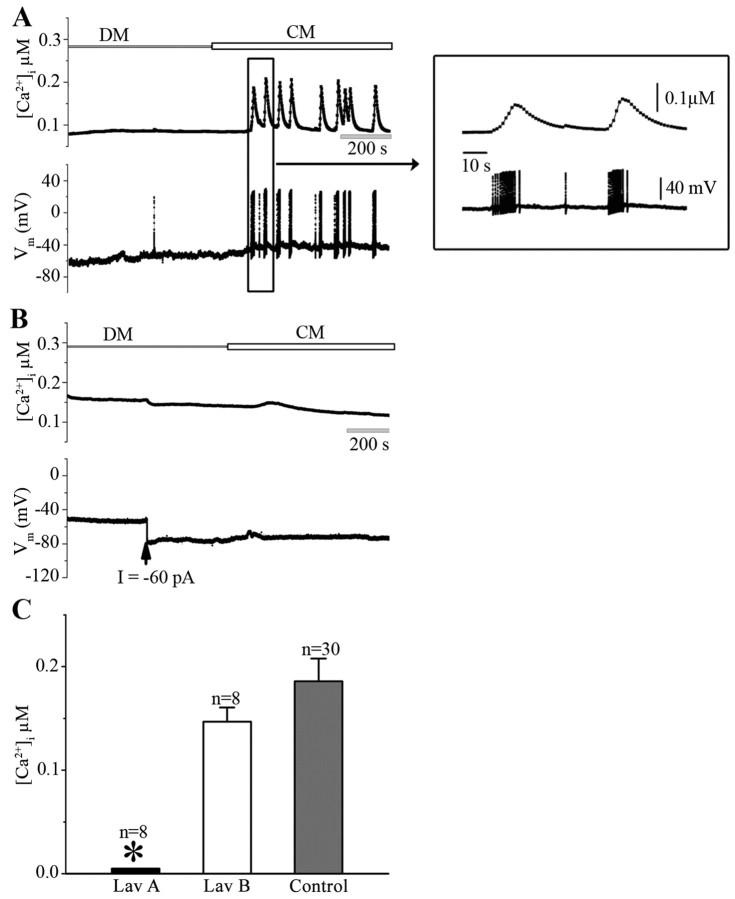
The CM-induced Ca2+ oscillations in LPeD1 neurons are activity-dependent and involve RTK activity. To determine whether CM-induced Ca2+ oscillations resulted from trophic factor-triggered action potentials, LPeD1 cells were cultured overnight in DM. On day 2, cells were loaded with Fura-2 AM and somata impaled with intracellular electrodes to monitor neuronal activity. Spontaneous action potentials were monitored in concomitant with ratiometric fluorescent images of LPeD1 first in DM and then in CM. A, Within minutes of CM exposure, LPeD1 neurons began to exhibit a bursting pattern and these bursts were almost always accompanied with Ca2+ oscillations, whereby [Ca2+]i raised from 138 ± 20 nm in DM to 286 ± 28 nm in CM (n = 10). The inset on right shows action potential bursts and Ca2+ activity on an extended time scale. B, To demonstrate that the observed Ca2+ oscillations indeed resulted from CM-induced burst of action potentials, LPeD1 neurons loaded with Fura-2 were first hyperpolarized by injecting −60 pA negative current (indicated by arrow) before neuronal exposure to CM. We found that neuronal hyperpolarization before CM exposure not only prevented action potentials but also the Ca2+ oscillations. C, To demonstrate that CM-induced increase in the intracellular Ca2+ involved RTK activity, LPeD1 cells cultured overnight in DM were first treated with RTK inhibitor Lav A (30 μm) and its inactive analog Lav B (30 μm). We found that Lav A, but not Lav B, significantly inhibited CM-induced rise in [Ca2+]i compared with the [Ca2+]i rise induced by CM alone (data from Fig. 3C). The asterisk indicates significant difference (t test; p < 0.05). Error bars indicate SE.

