
Figure 4.
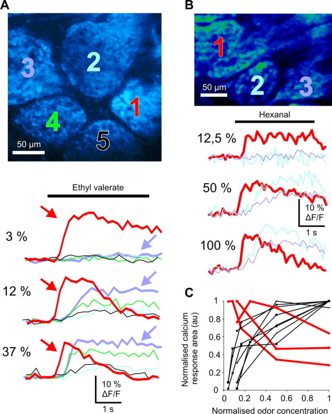
Peripheral adaptation causes a spatial redistribution of activated glomeruli. A, B, Top, Two-photon imaging of two large fields of view comprising five (A) and three (B) glomeruli. ORN terminals were labeled with Ca2+ green dextran (10 kDa). A, Bottom, Glomeruli presented two types of Ca2+ responses according to the odorant concentration. At low concentration, ethyl valerate evoked a rapid Ca2+ plateau (red arrow) in glomerulus 1, although it did not affect neighboring glomeruli (2–5). Raising the odorant concentration shortened the Ca2+ response of glomerulus 1, although it revealed those of glomeruli 2–5. Note the appearance of a Ca2+ plateau in glomerulus 2 (blue arrow). B, Bottom, Another case where the most sensitive glomerulus (glomerulus 1) shows a Ca2+ response adaptation. C, Quantification of Ca2+ signal adaptation. The overall Ca2+ responses (area) were normalized and plotted as a function of the odorant concentration (normalized to its maximum value) for three groups of glomeruli (11 glomeruli, three animals). Peripheral adaptation greatly reduced Ca2+ responses of the three most sensitive glomeruli (red traces), whereas responses of the neighboring glomeruli increased (black traces).