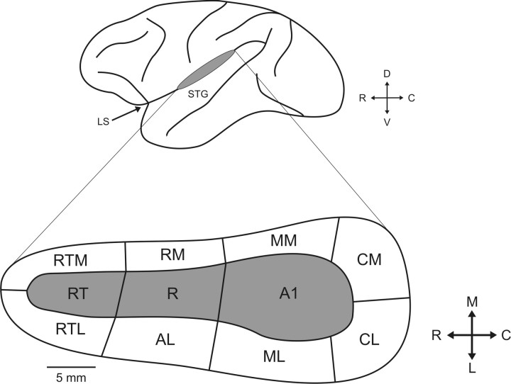
Figure 1.
Schematic of the lateral view of the macaque brain (above), with the location of auditory cortex within the lateral sulcus indicated by a gray ellipse. The lower inset is a schematic identifying the three core (gray) and eight belt (white) regions. LS, Lateral sulcus; STG, superior temporal gyrus; RT, rostrotemporal field; AL, anterolateral area; RTL, lateral rostrotemporal area; RTM, medial rostrotemporal area; RM, rostromedial area; MM, middle medial area; D, dorsal; V, ventral; M, medial; L, lateral.

