
Figure 6.
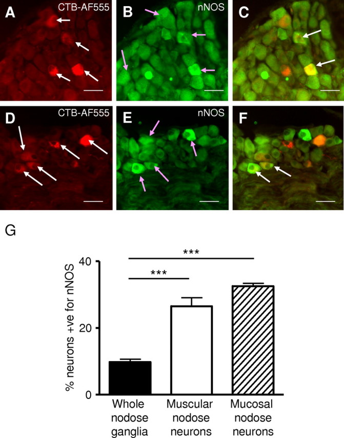
Combined nNOS immunohistochemistry and retrograde tracing in the mouse nodose ganglia. A, Mouse nodose ganglia showing retrogradely traced (CTB-AF555) gastric mucosal afferent neurons (red; indicated by the white arrows). B, Neurons immunoreactive for nNOS (green; indicated by pink arrows). C, nNOS immunoreactivity (green) and colabel (yellow; indicated by white arrow). Two gastric mucosal afferents are seen prominently colabeled with nNOS (indicated by white arrows). D, Retrogradely traced (CTB-AF555) gastric muscular afferent neurons (red; indicated by white arrows). E, Neurons immunoreactive for nNOS (green; indicated by pink arrows). F, nNOS immunoreactivity (green) and colabel (yellow; indicated by white arrow). Two gastric muscular afferents are prominently colabeled with nNOS (indicated by white arrow). Scale bars, 150 μm. G, The percentage of neurons in the nodose ganglia immunoreactive for nNOS. The percentage of neurons immunoreactive for nNOS in the whole nodose ganglia is significantly less (***p < 0.001; unpaired t test) than the percentage of both gastric mucosal and muscular afferent neurons immunoreactive for nNOS.