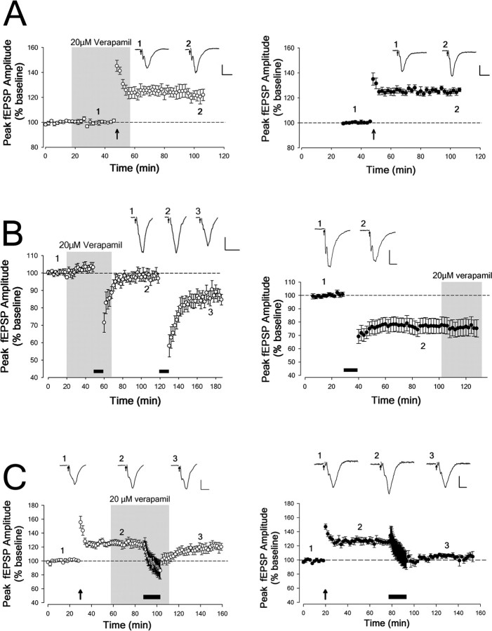
Figure 5.
Effects of verapamil on synaptic plasticity in PRH slices. The presence of verapamil in the perfusate is indicated by a gray box. The traces illustrate fEPSPs taken at the time points indicated. Stimulus artifacts are blanked and replaced by arrows. Calibration: 0.2 mV, 10 ms. A, LTP (induced by 100 Hz HFS repeated 4 times, as indicated by the upward arrow) was readily induced in the presence of 20 μm verapamil (left; open circles). LTP induced in the presence of verapamil was not different from that induced in control slices (right; closed circles). B, De novo LTD (induced with 5 Hz stimulation indicated by the filled bar) was prevented in the presence of 20 μm verapamil; after washout, delivering an additional train of 5 Hz stimulation resulted in significant LTD (left; open circles). Verapamil had no significant effect on the expression of LTD, which was tested by bath applying 20 μm verapamil 60 min after induction of LTD in control slices (right; closed circles). C, After induction of LTP, depotentiation (induced by 1 Hz stimulation for 15 min as indicated by the filled bar) was also prevented by 20 μm verapamil (left; open circles). This contrasts with control experiments, in which 1 Hz LFS resulted in reversal of LTP to baseline (right; closed circles).

