
Figure 6.
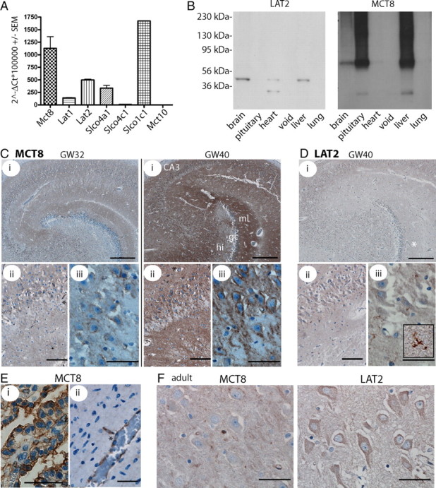
Developmental expression of thyroid hormone transporters in the human brain. A, qPCR detection of thyroid hormone transporters in adult human brain cDNA. Values are calculated according to the ΔCt method in relation to β-actin as a housekeeping gene. B, Multiple tissue Western blot on adult human membrane fractions for LAT2 and MCT8. The antibodies detect a protein of the same size as in mouse brain. Left, Molecular weight markers. C, Immunohistochemical detection of MCT8. Labeling of MCT8 increases with time in the developing human brain. i, Hippocampus. Scale bar, 500 μm. ii, CA3. Scale bar, 100 μm. iii, CA3. Scale bar, 50 μm. GW32 and GW40 are shown. D, Immunohistochemistry for LAT2 in GW40 hippocampus detects microglial, but not neuronal staining (iii, inset). Scale bars are as in C. E, MCT8 in GW25 choroid plexus (i) (scale bar, 50 μm) and cortical gray matter vessel (ii) (scale bar, 50 μm). F, Hippocampal CA3 neurons in the adult human brain stained for MCT8 and LAT2. Scale bars, 50 μm. Capillaries are indicated by asterisks.