
Figure 2.
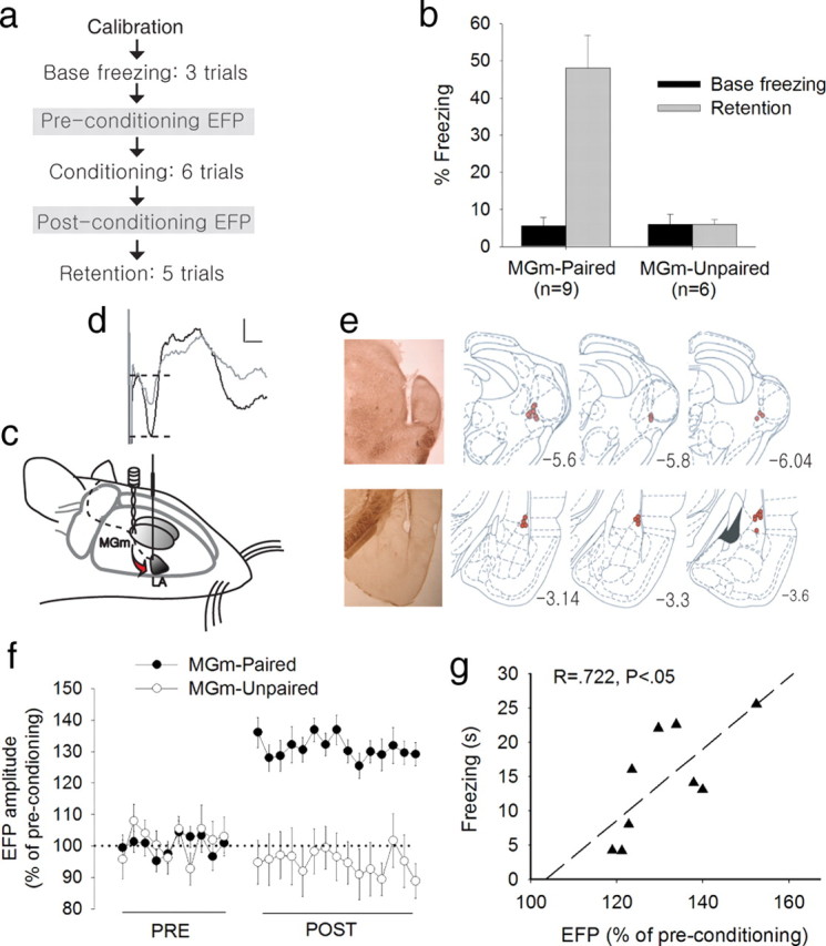
Enhanced EFP in the LA after fear conditioning. a, Experimental procedure for experiment 2. The procedure is identical to experiment 1 except that the two EFP sessions were inserted before and after conditioning. b, Changes in freezing during the retention test. Only the MGm-paired group showed significant freezing to the CS compared with the preconditioning baseline. c, Arrangement of stimulating and recording electrodes in the brain for fear conditioning and LA recording. d, A representative EFP waveform on a single trial (gray line, preconditioning; black line, postconditioning). EFP amplitude was defined as the difference between the first positive peak and the first negative valley, marked by two dotted horizontal lines. Calibration: 200 μV, 5 ms. e, Electrode placement. Photos show stimulating (top) and recording (bottom) sites, marked by small electrolytic lesions at the tip. All of the stimulating electrodes were placed in the MGm or its immediate vicinity. All recording electrodes were found in the dorsal subdivision of the LA. The drawings were modified from Paxinos and Watson (1998). f, Average EFP data showing percentage changes after conditioning. Each data point represents an average of two trials presented at every 30 s. The MGm-paired group showed significantly enhanced EFP compared with the preconditioning baseline, whereas the MGm-unpaired group showed no significant difference. In addition, the MGm-paired group also showed greater EFP than the MGm-unpaired group during the postconditioning test. g, Relation between fear memory retention and postconditioning EFP enhancement.