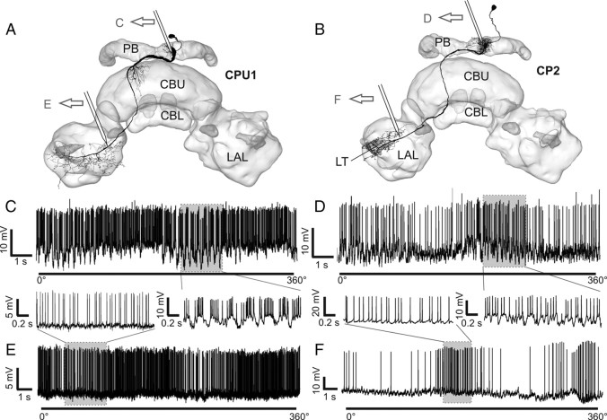
Figure 6.
Indication of presynaptic and postsynaptic arborizations by recordings from different regions of CPU1 and CP2 neurons. A, B, Frontal reconstructions of a CPU1 (A) and CP2 neuron (B), projected onto a three-dimensional reconstruction of the CC. Approximate recording sites of the recording traces in C–F are indicated. C, Recording trace from arborizations of a CPU1 neuron in the PB during zenithal rotation of the polarizer. Postsynaptic, graded potentials are visible (enlargement of shaded area is shown in the inset). D, Recording trace from PB-arborizations of a CP2 neuron during a rotation of the polarizer. Postsynaptic potentials are clearly visible (enlarged in the inset). E, F, Recording traces obtained from the vicinity of the LAL from a CPU1 neuron (E) and a CP2 neuron (F). Enlargements emphasize the even baseline without major graded potential changes. LT, Lateral triangle. Neuron morphologies in A and B are modified from Heinze and Homberg (2008).

