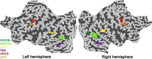
Figure 2.
Analyzed regions of interest. Regions of interest for a single subject visualized on the flattened cortical surface of the left and the right hemisphere of this subject. The color coded areas represent, the human MT complex (hMT/V5+), the kinetic occipital area (KO/V3b), the pSTS, the FBA, and action responsive regions in the vPrCS, and the pIFS. White lines delineate early visual areas V1 and V2. Dark gray, Sulci; light gray, gyri. prCS, Precentral sulcus; CS, central sulcus; STS, superior temporal sulcus; IPS, interparietal sulcus; OTS, occipital temporal sulcus.

