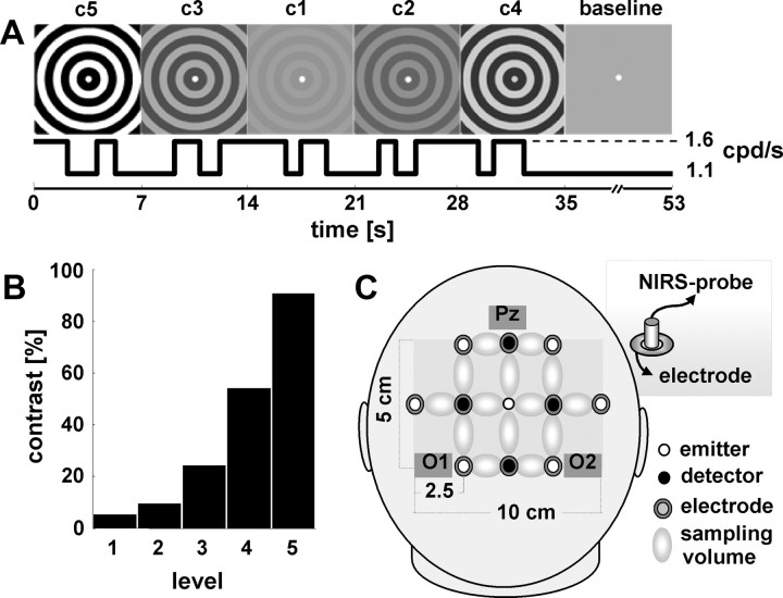
Figure 1.
Experimental design. A, Concentrically moving gratings changed contrast (c1–c5) every 7 s in a pseudorandomized order. Within each 7 s period, the subject had to detect and respond to three changes in velocity, which alternated between 1.1 and 1.6 cpd/s. Because contrast and velocity were modulated independently, external modulation of stimulus salience (contrast) and additional internal response determinants could be disentangled by means of reaction time. An 18 s baseline period, for which contrast was kept at 0%, was inserted every 35 s (i.e., after 5 contrast changes). B, Michelson contrasts of the gratings tested (c1–c5). C, Sketch of the probe arrangement over the occipital-parietal region. Optical probes were inserted into electrode rings (inset), allowing for simultaneous measurements of electrophysiological and oxygenation response over the same area.

