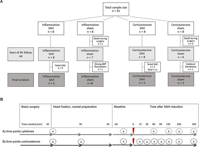
Fig 2. Experimental design.
A: Diagram of sample size distribution: 32 animals were randomly allocated to four different groups for blood sampling for inflammation markers (IL-6 and TNF-α) and corticosterone; after subtracting drop-outs from surgery and recording phase, a total of 21 animals were included in the final analysis. Subarachnoid hemorrhage (SAH) was induced by injection of 500μl arterial blood within 1 minute into the cisterna magna, sham animals did not receive any injection. B: Time points for blood sampling for inflammatory cytokine and corticosterone analysis: The diagram shows the time points (x) when blood was withdrawn within basic surgery (anaesthesia, tracheotomy, ventilation, A./V. femoralis cannulation), head fixation and cranial preparation (turning to prone position, fixation with ear bars in stereotactic frame, cranial window preparation, trepanations for EEG and ICP recording, cannula for icv blood injection) followed by the measurement period; red flash marks the time point of SAH induction.

