
Fig. 2.
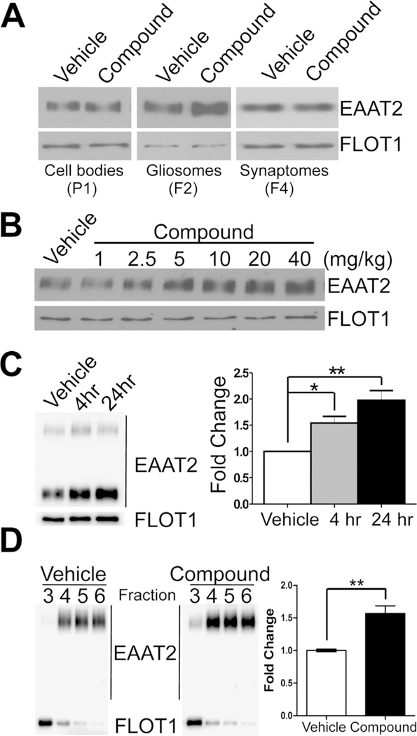
Pyridazine analogs specifically and rapidly increase EAAT2 expression only in the PAP. Mice were treated with compound at 40 mg/kg (or as depicted) and forebrains were harvested for the gliosome preparation 24 h (or as described) post-treatment. (A) Western blots show robust increases of EAAT2 protein expression were only found in gliosomes (F2) while no induction was observed in the cell bodies (P1) or in synaptosomes (F4). (B) Dose-dependent increase in EAAT2 protein expression in response to single, escalating compound dose. (C) Time-dependent fold increase in EAAT2 protein expression in response to compound treatment at 4 h (1.54 ± 0.13) and 24 h (1.98 ± 0.19) post-treatment. Quantification of EAAT2 expression time course (normalized to flotilin-1 protein; n = 4/group). (D) EAAT2 expression increased in the lipid raft micro-domain, which represents the functional EAAT2 lipid domain, of the forebrain (1.57 ± 0.12) after a single treatment. Data represented as mean ± SEM and analyzed using a one-way ANOVA with Tukey’s post-hoc test. Statistical significance denoted by *p < .05; **p < .01.