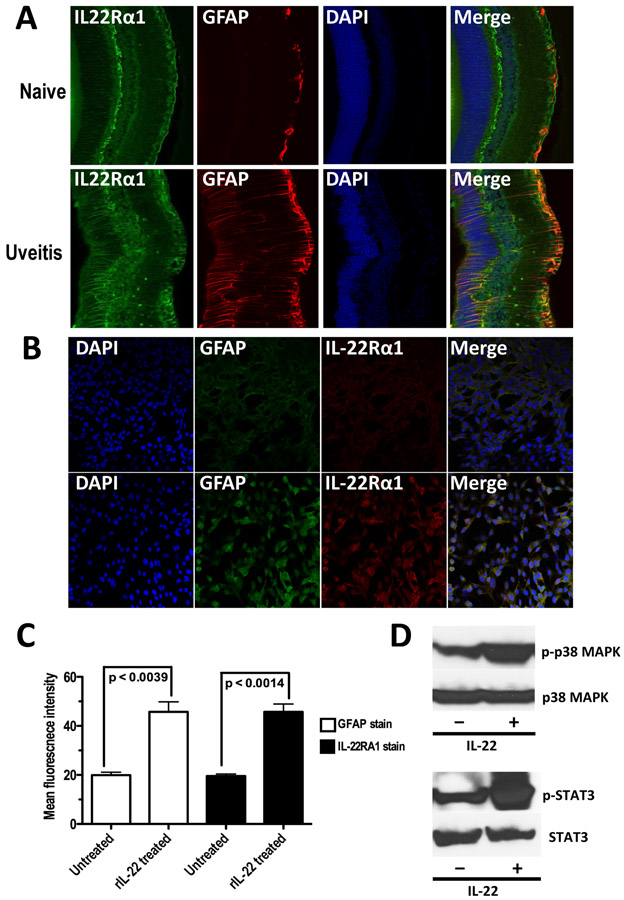
Figure 4: Retinal Muller cells express IL-22R α1 and respond to IL-22 stimulation by activating Stat3 and Mapk.
(A) Retinal sections (thickness of 100μm using Vibratome) of eyes from naive and IRBP immunized (day 22) C57BL/6J mice were stained with anti-human IL-22Rα1 (green), anti-mouse GFAP (red), and DAPI. Images from naive eye and uveitic eye are shown. (B) Expression of IL-22Rα1 was enhanced when Muller cells in culture was treated in-vitro with rIL-22. Adherent Muller cells were stained with DAPI, anti-mouse GFAP (Alexa 488), and anti-human IL-22Rα1 (Alexa 555). Images from untreated Muller cells and rIL-22 treated (100ng/ml for 24 hours) Muller cells are shown. (C) Mean fluorescent intensity of GFAP and IL-22Rα1 staining in rIL-22 treated and untreated Muller cells show significantly high receptor expression within 24 hours. p-value is calculated by two-tailed unpaired ‘t’ test. (D) Phosphorylation of Stat3 and p38 subunit of Mapk was enhanced in Muller cells when treated with rIL-22 (500ng/ml for 45 minutes) compared to that in untreated Muller cells.

