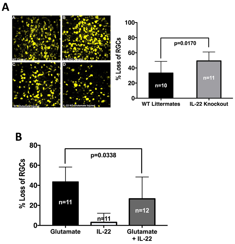
Figure 7: Presence of IL-22 protects Retinal Ganglion Cells from glutamate induced neurotoxicity.
C57BL/6J, IL-22 KO or its wild type littermates were subjected to retrograde labeling of RGCs with fluorogold followed by single intravitreal injection of glutamate with or without rIL-22. (A) Representative retinal flat mount images (40X magnification) from WT littermates or IL-22 KO mice with or without glutamate injury are shown. Histogram shows percentage loss of RGCs normalized to the contralateral control eyes from two independent experiments. IL-22 KO mice experienced significantly higher loss of RGCs when subject to glutamate injury. Data were analyzed by Welch’s unpaired ‘t’ test for significance. (B) Fluorogold labeled C57BL/6J mice were injected intravitreally with glutamate alone, rIL-22 alone or glutamate + rIL-22. Loss of RGCs in each group was normalized to a control group intravitreally injected with PBS. Data from two independent experiments were pooled and analyzed using one way ANOVA. Presence of IL-22 significantly decreased the loss of RGCs due to glutamate toxicity.

