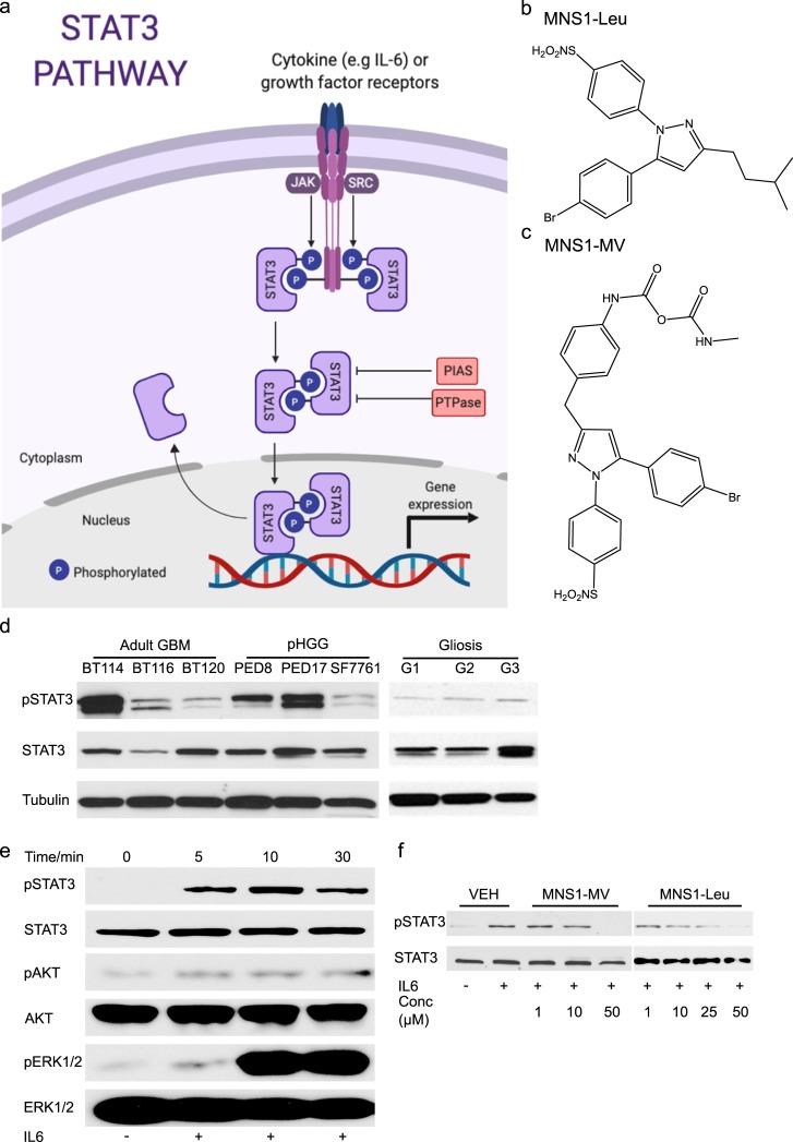
Fig 1. Pyrazole-based MNS-1 inhibitors block STAT3 phosphorylation.
(a) Diagram of STAT3 pathway; activation is by cytokines, most commonly IL-6, and growth factor receptors, most commonly gp130. Phosphorylated STAT3 forms a reciprocal homodimer that translocates into the nucleus where it interacts with DNA and activates gene transcription. The activated form is typically inhibited by Protein inhibitor of activated STAT (PIAS) and Protein-tyrosine Phosphatase (PTPase). (b) Chemical structure of MNS1-Leu. (c) Chemical structure of MNS1-MV. (d) Western blot of pSTAT3 (Y705) in adult GBM and pHGG patient derived cell lines at baseline compared to non-tumor brain control (removed for epilepsy). (e) Western blot analysis of IL-6 (100 ng/mL) stimulation of STAT3 phosphorylation in dBT114 for 5–30 min. (f) Western blot analysis on the effect of IL-6-induced STAT3 phosphorylation in dBT114 in presence of MNS1-MV and MNS-Leu in various concentrations after a 10 min exposure to IL-6 stimulation.

