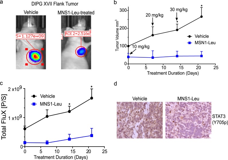
Fig 5. MNS1-Leu slows pHGG growth in vivo and reduces STAT3 activation.
Dose escalation study with MNS1-Leu in PDX model of pHGG (DIPG17) starting at 10 mg/kg and increasing to 30 mg/kg over 21 days. (a) BLI imaging in PDX model of vehicle control vs. MNS1-Leu at day 21. (b) Caliper measurements of flank “DIPGXVII” xenograft tumors showed significant increase in tumor size when treated by vehicle, but not when treated by MNS1-Leu. (c) Similarly, bioluminescence of these tumors also showed significantly increased flux in those treated by vehicle, with no difference in those treated by MNS1-Leu. Values are the means ± S.E.M (error bars) of triplicate experiments. (d) Immunohistochemistry staining for pSTAT3 in these tumors demonstrated a decrease in pSTAT3 expression in those treated by MNS1-Leu compared to those treated by vehicle.

