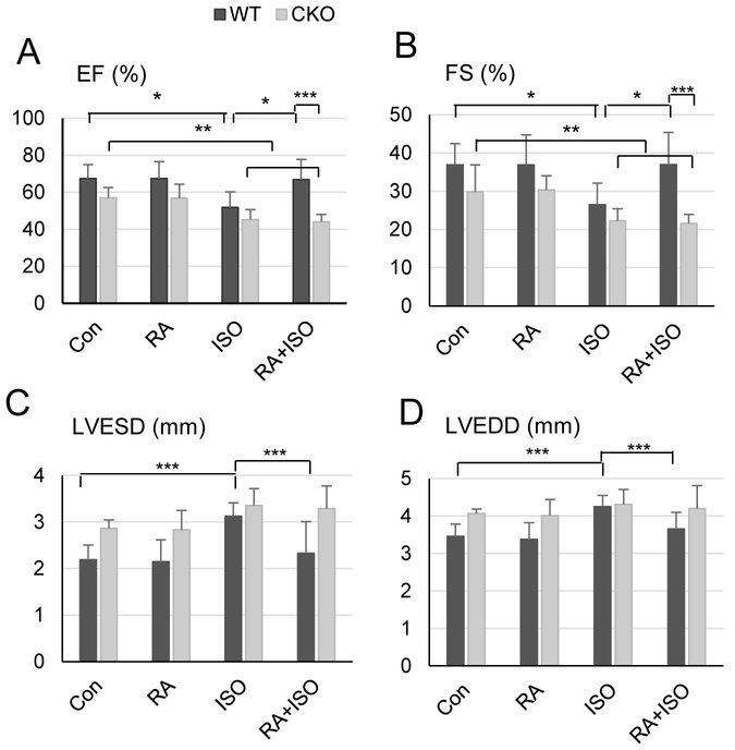
Fig. 1.
RA prevents cardiac dysfunction in WT, but not CKO, mice induced by β-adrenergic stimulation. M-mode echocardiography was performed on the WT and CKO mice pre-treated with RA and then ISO for 7 d at the age of 16-24 weeks (n = 5-10). Data were expressed as means ± SDs. Ejection fraction (EF, panel A) and fractional shortening (FS, panel B) represent cardiac function. Left ventricular end-systolic dimension (LVESD, panel C) and left ventricular end-diastolic dimension (LVEDD, panel D) represent internal diameters of left ventricle. *P < 0.01, **P < 0.05, ***P < 0.001 (two-way ANOVA, Student-Newman-Keuls method).

