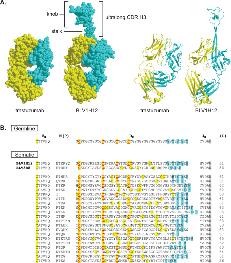
Figure 1.
Structure and diversity of cow ultralong CDR H3 regions. (A) The Fab fragments for trastuzumab (PDB: 1N8Z) and BLV1H12 (PDB: 4K3D) are shown in space fill (left) and ribbon diagrams (right). The heavy chains are colored cyan and the light chains yellow. The ultralong CDR H3 in the cow Fab BLV1H12 is comprised of a β-ribbon “stalk” and disulfide bonded “knob” domain that protrude far from the typical antibody paratope. (B) Sequences of ultralong CDR H3s are shown, with the germline VHBUL, DH2 and JH illustrated at the top and several somatic sequences at the bottom. The sequences encoding the two Fabs whose crystal structures have been solved (BLV1H12 and BLV5B8) are indicated. Cysteines are highlighted in yellow, with those encoded by the germline DH2 underlined and in red. Aromatic residues expected to form the descending strand of the β-ribbon stalk are in cyan, and the conserved tryptophan in the JH region is greyed. Note the conservation of the first cysteine encoded by DH2, with the decrease in conservation proceeding C-terminally.

