Fig. 3.
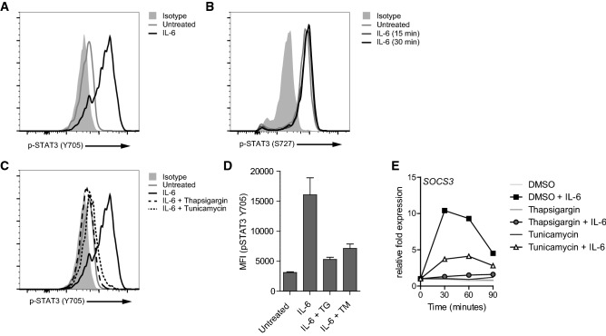
ER stress-induced suppression of STAT3 phosphorylation is not restricted to IL-10 signaling. Macrophages were stimulated with 25 ng/mL IL-6 for 15 min (a, b) or 30 min (b) and analyzed for phosphorylation of STAT3 at Tyr705 (a) and Ser727 (b) using flow cytometry. Light grey histogram indicates background staining. c, d Macrophages were pre-treated with thapsigargin (TG), tunicamycin (TM), or vehicle control and stimulated with 25 ng/mL IL-6 for 15 min. Phosphorylation of STAT3 at Tyr705 was analyzed using flow cytometry. Light grey histogram indicates background staining. Representative example (c) and pooled MFI data (D) from three different experiments, mean + SEM. e Macrophages were pre-treated with TG, TM or vehicle control and stimulated with 25 ng/mL IL-6 and analyzed for mRNA expression of SOCS3 using qPCR (normalized to GAPDH expression, fold increase compared to unstimulated control). Data (a–d) are representative examples of three independent experiments using different donors

