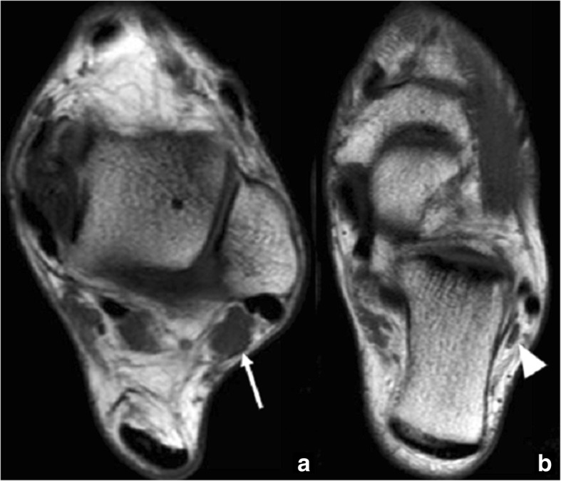
Fig. 10.

Peroneus quartus. 44-year-old man referred for follow-up of an osteochondral lesion in the talus. Axial FSE T1 images in different planes (a) proximal and (b) distal demonstrate the incidental finding of a peroneus quartus (white arrow). This descends to insert in the lateral aspect of the calcaneus, with a fleshy attachment (arrowhead)
