Fig. 3.

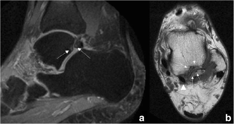
52-year-old man with persisting posterior ankle pain. a Sagittal fast spin-echo proton density (FSE PD) fat sat demonstrates an os trigonum. There are foci of increased signal intensity in the subchondral bone in both aspects of the synchondrosis (white arrows), with a small amount of fluid in the posterior aspect of the tibio-talar joint. b Axial fast spin-echo T1 (FSE T1) better depicts the presence of foci of subchondral bone oedema and subchondral bone cysts in both aspects of the synchondrosis (white arrows). Note how the os trigonum is intimately related to the flexor hallucis longus (arrowhead)
