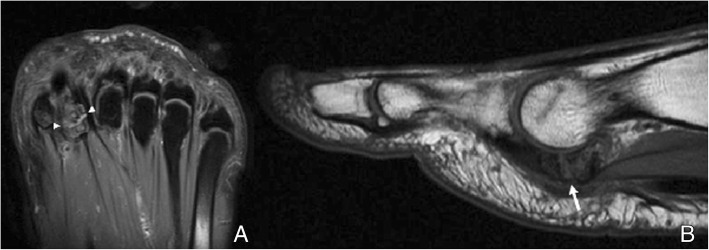
Fig. 13.
Lateral sesamoid fracture. A 35-year-old man with pain over plantar aspect of foot after trauma. a Axial FSE PD fat sat demonstrates a fracture of the lateral sesamoid, with slight separation of fragments, and a band of interposed fluid (white arrowheads). b Sagittal FSE T1 demonstrates hypointensity in the fracture fragments. The fracture line is visible, slightly more hyperintense than the adjacent fragments (white arrow)

