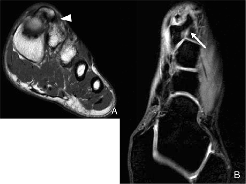
Fig. 18.

Os intermetatarseum. A 36-year-old man, investigated for pain in the Achilles. Radiographs demonstrate a spur projection in the dorsum of the foot, overlying the base of the metatarsal (not shown). a Coronal FSE T1 demonstrates how this articulates with the base of the second metatarsal, with a synchondrosis (white arrowhead). b Axial three-dimensional gradient echo water selective/fluid (WATSf) demonstrates this exostosis arises from the base of the first metatarsal, and extends over the joint with the base of the second metatarsal (white arrow). These ossicles rarely represent a cause or pathology, unless compression irritates the superficial and deep peroneal nerves
