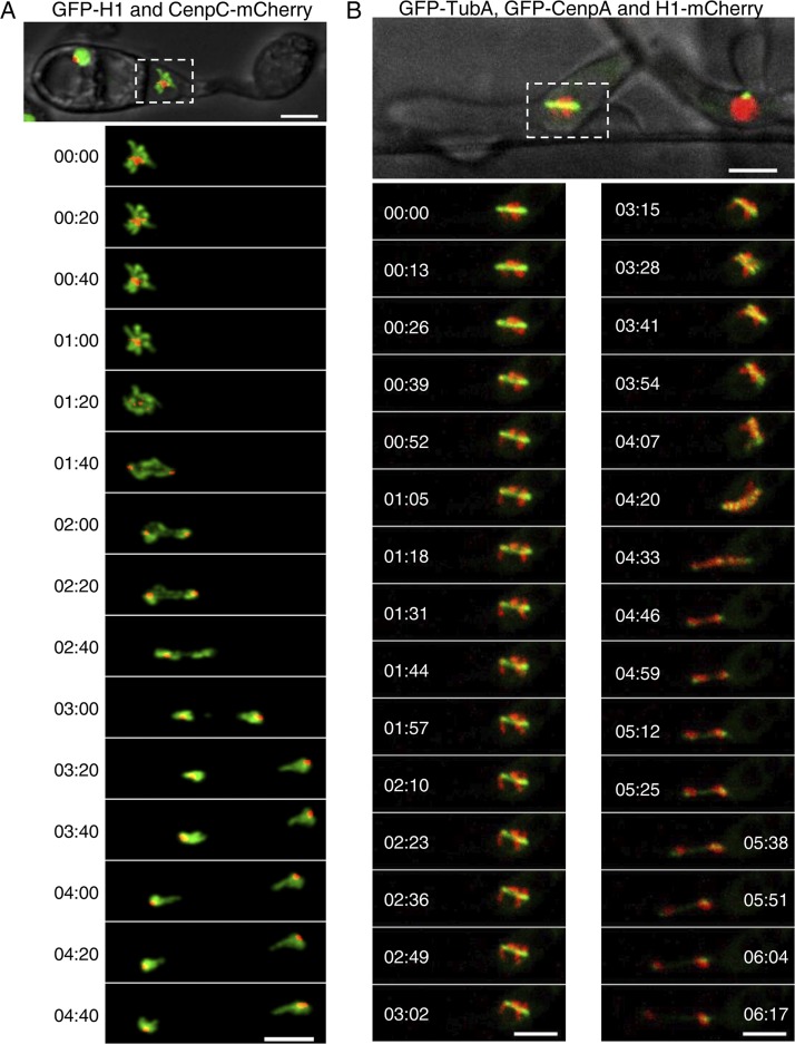
FIG 3.
Subcellular localization and dynamics of CenpA and CenpC during pathogenic development in M. oryzae. (A) Time-lapse images showing a mitosis event during appressorium formation in CenpC-mCherry and GFP-histone H1-tagged strain B157 of M. oryzae. Conidia were incubated on the hydrophobic coverslip to allow appressorium development, and the mitotic division was recorded after 4 h postinfection (hpi), with images captured at 20-s intervals (see also Movie S5 at https://figshare.com/articles/MoCEN_movies/8282066). (B) Time-lapse images showing the localization of centromeres (GFP-CenpA), microtubules (GFP-TubA), and the nucleus (histone H1-mCherry) during mitosis in the invasive hyphae in M. oryzae. M. oryzae conidia were incubated on rice sheath, and the images were acquired at 44 hpi at 13-s intervals (see also Movie S6 at https://figshare.com/articles/MoCEN_movies/8282066). The epifluorescent confocal images shown here are maximum projections from Z-stacks consisting of 0.5-μm-spaced planes. Bars, 5 μm.

