
FIGURE 2.
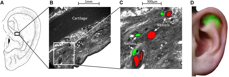
Wiring of vessels and nerves in the ear for the percutaneous aVNS. (A–C) High-resolution episcopic images of a volume biopsy in the cymba conchae of one male cadaver ear. Indicated blood vessels (in red) and nerves (green) reside apparently close to each other indicating their joint proliferation in the ear. (D) In order to find local auricular nerve branches, the outer ear is transilluminated to localize and visualize easily discernable auricular vessels which are less transparent than the surrounding tissue for green light. The visualized locations of vessels indicate the most likely regions of nerves, which serve for a personalized placement of stimulation needles.