Figure Legend 1:
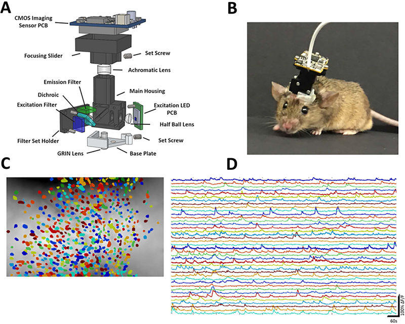
Calcium imaging of neuronal activity in freely moving mice.
A. Schematic demonstrating all the components of the open-source miniaturized microscope. B. Photograph of mouse walking with a miniaturized microscope imaging the hippocampus. C. Schematic demonstrating a subset of the neurons imaged from hippocampal CA1 in freely behaving mice. D. Calcium traces from neurons demonstrated in C.

