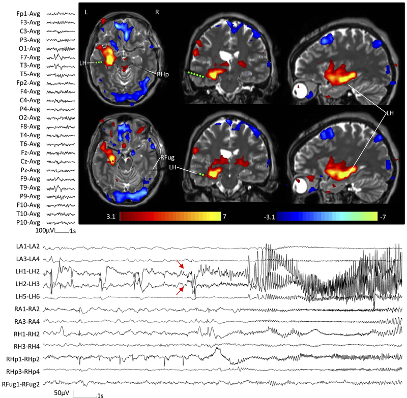
Figure Legend 4:
EEG-fMRI findings in temporal lobe epilepsy.
In this example, EEG-fMRI hemodynamic responses (top right) correlated with epileptic discharge over the left temporal region on scalp EEG (top left). The hemodynamic response involved a widespread network, with the maximum in the epileptogenic zone (in this case the left hippocampus). The patient underwent a stereo-encephalography study, which confirmed the left hippocampus as the main generator of the seizures (bottom). Red arrows indicate the EEG onset of the seizure.

