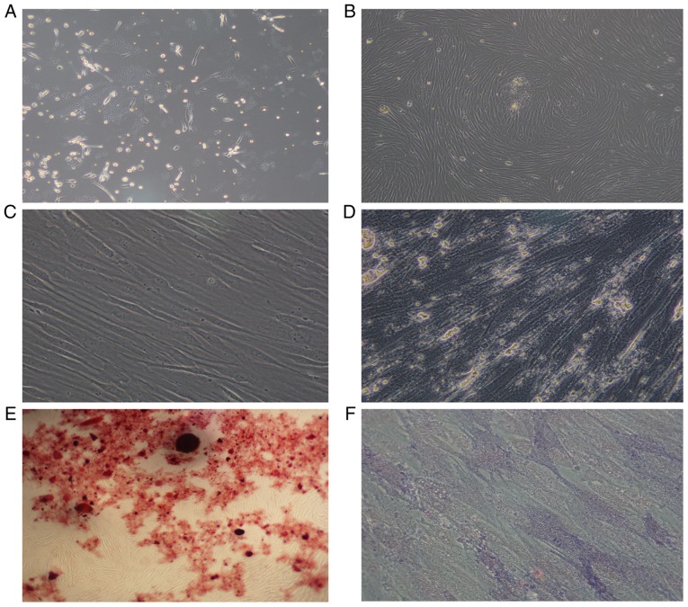
Figure 1.
Morphology of AML-MSCs. (A) AML-MSCs cultured after 7–10 days. (B) AML-MSCs cultured after 14–21 days. (C) Subculture of AML-MSCs. (D) AML-MSCs osteogenic induction culture for 14 days. (E) AML-MSCs stained with alizarin red. (F) AML-MSCs stained with alkaline phosphatase. Magnification was as follows: (A) ×10; (B) ×4; (C) ×40; (D) ×40; (E) ×4; (F) ×40. AML-MSCs, acute myeloid leukemia derived-mesenchymal stem cells.

