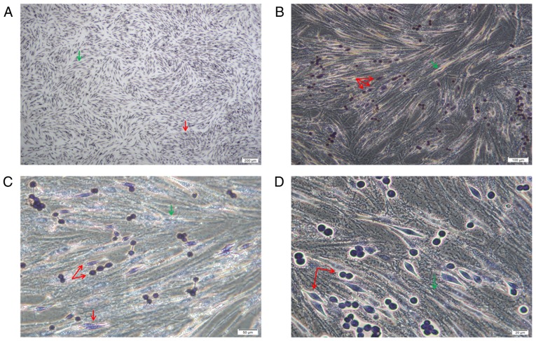
Figure 3.
(A-D) POX staining. The bottom spindle-shaped cells were AML-MSCs negative for POX staining (green arrows). The superficially adherent K562-ADM cells had two forms, one was a normal spherical cell, and the other was a spindle-shaped cell. All cells were stained positive for POX (red arrows). Magnification was as follows: (A) ×4; (B) ×10; (C) ×20; (D) ×40. AML-MSCs, acute myeloid leukemia derived-mesenchymal stem cells.

