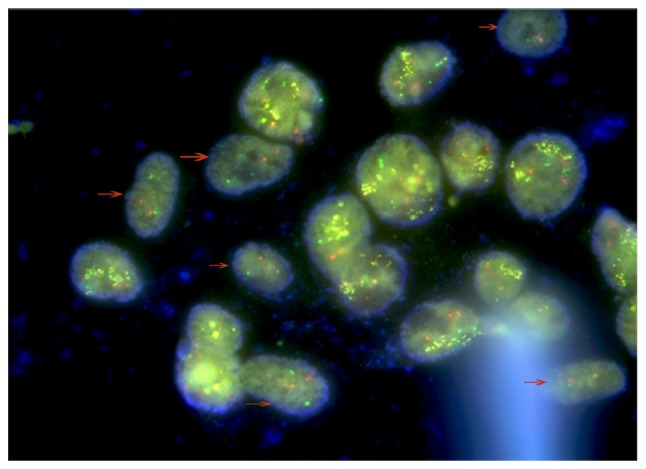
Figure 5.

FISH analysis. FISH assay revealed that there were 2 types of cells in the visible field. The cells indicated by the arrows are AML-MSCs with a negative BCR/ABL expression. Other cells are the K562-ADM cells which were positive for BCR/ABL expression (49). FISH, fluorescence in situ hybridization; AML-MSCs, acute myeloid leukemia derived-mesenchymal stem cells.
