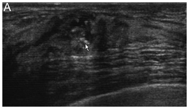
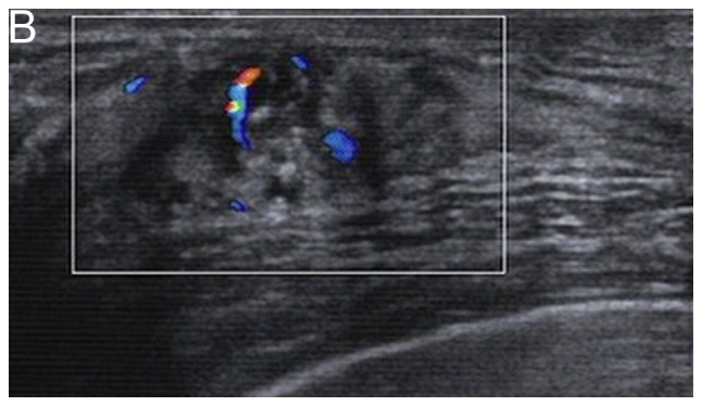
Figure 1.


A 43-year-old premenopausal woman presented with a palpable right breast mass and a diagnosis of ductal carcinoma in situ with microinvasion. (A) A grayscale sonogram demonstrates a hypoechoic echo non-mass abnormality. Punctate echogenic foci within the lesion represent associated calcifications (arrow). (B) A color Doppler sonogram shows the presence of vascularity.
