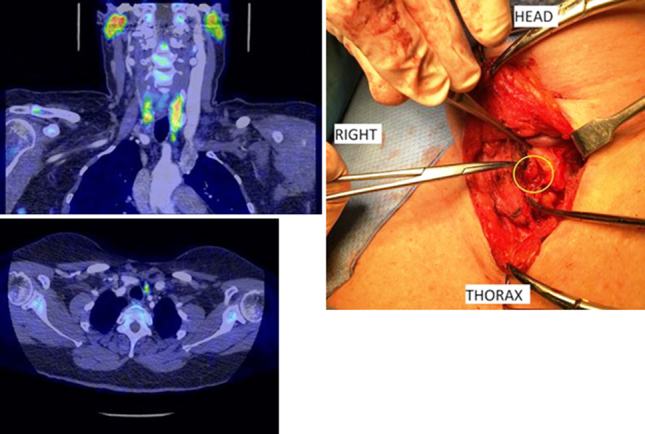
Figure 2.

A 59-year-old woman with four previous failed neck explorations. The 18fluorocholine positron emission tomography/computed tomography (PET/CT) scan correctly identified a 5-mm mildly choline-avid focus (maximum standardised uptake value 4.1) immediately abutting the inferior pole of the left lobe of thyroid, intimately related to a small vessel, which was removed and the patient cured (axial and coronal fused PET/CT images on a standardised uptake values 0–6 scale using the arterial enhanced CT images and intraoperative picture).
