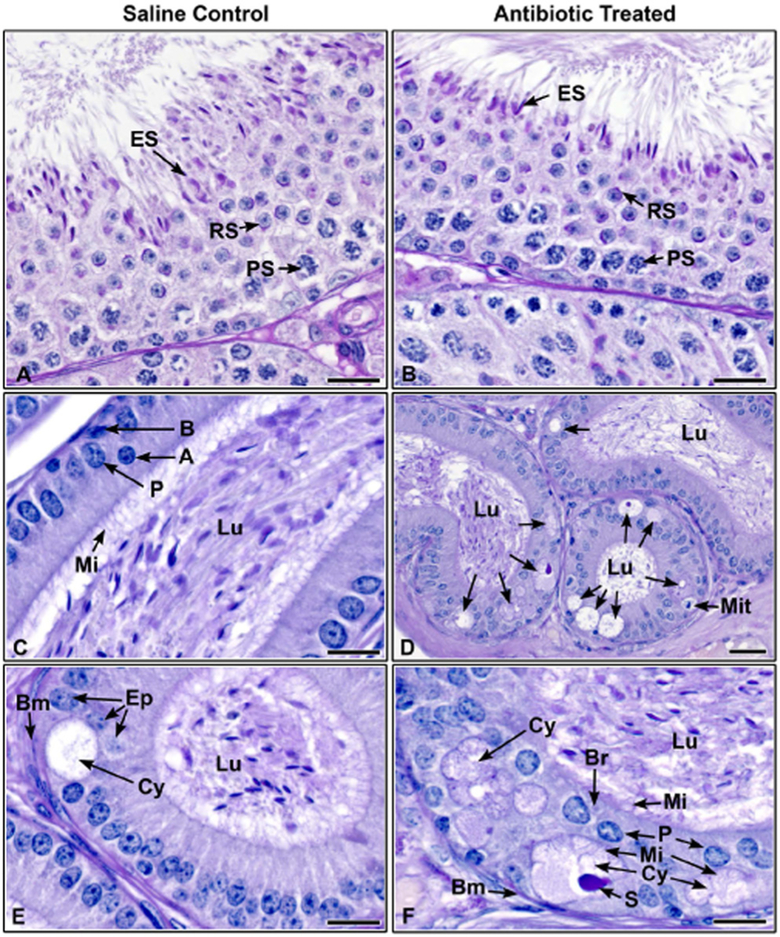
Figure 6.
Histological section of testes and epididymides from saline control and CUB antibiotic-treated males. (A) Testis from saline control male showing normal spermatogenesis, Stage VII. Elongated spermatids (ES) line the epithelium with tails extending into the lumen. Round spermatids (RS) with normal acrosomal formations, as seen with periodic acid-Schiff’s staining (PAS) are found in the central region of the epithelium (PS, pachytene spermatocytes). (B) Testis from CUB antibiotic-treated male, late Stage VII. Normal elongated spermatids (ES) line the epithelium as in the control. Round spermatids (RS) with normal acrosomal formations are present, as well as pachytene spermatocytes (PS). (C) Proximal corpus epididymis from a control male. Note the concentration of sperm in the lumen (Lu) and a columnar epithelium consisting of principal (P), apical (A) and basal (B) cells. Long microvilli (Mi) extend into the lumen. (D) Proximal corpus epididymis from a CUB antibiotic-treated male showing cribriform cystic changes (arrows) in the epithelium. These abnormal formations appear as large vacuoles in the basal region that develops into a cystic luminal space, surrounded by shortened epithelial cells. Sperm are found in the epididymal lumen (Lu). A mitotic figure (Mit) is seen in the basal region. (E) Proximal corpus epididymis from a control male showing a rare cribriform cyst in the epithelium. This cyst (Cy) appears less developed than in the treated samples, because there is a sparse amount of secretory material and microvilli are lacking within the cystic lumen (Bm, basement membrane; Lu, lumen of the epididymis). (F) CUB antibiotic-treated proximal corpus epididymis. This his higher magnification of an area from D, showing more well-developed cribriform cysts (Cy), with some showing epithelial cells underneath on the basement membrane (Bm), as well as a well-developed ‘bridge’ of epithelial cells (Br). These cysts have well-defined secretory material (S) in the cystic lumen and the lining epithelial cells show microvilli (Mi) extending into both the cyst as well as the lumen (Lu) of the epididymis. Bars = 20 μm.

