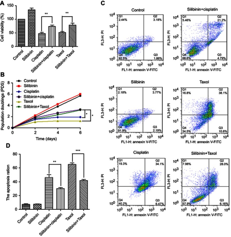
Figure 5.
Silibinin reduce cisplatin-induced and taxol-induced hepatotoxicity. (A) The LO2 cells were treated with silibinin (50 μM), cisplatin (24 μM), taxol (4 μM) and silibinin (50 μM) plus cisplatin (24 μM) and/or taxol (4 μM) for 48 h, and then the cell viability was determined by MTT assay. The results were shown as the percentage of cell viability in control group. (B) Proliferation curve of LO2 cells in the presence of silibinin (50 μM), cisplatin (24 μM), taxol (4 μM) and silibinin (50 μM) plus cisplatin (24 μM) and/or taxol (4 μM). (C) silibinin (50 μM), cisplatin (24 μM), taxol (4 μM) and silibinin (50 μM) plus cisplatin (24 μM) and/or taxol (4 μM) treatment induces apoptosis of LO2 cells. Apoptotic cells were assayed by Annexin V/PI staining and FACS analysis. (D) Quantification of (C). Values are the average ± SD of three independent experiments. *p<0.05, ***p< 0.001.

