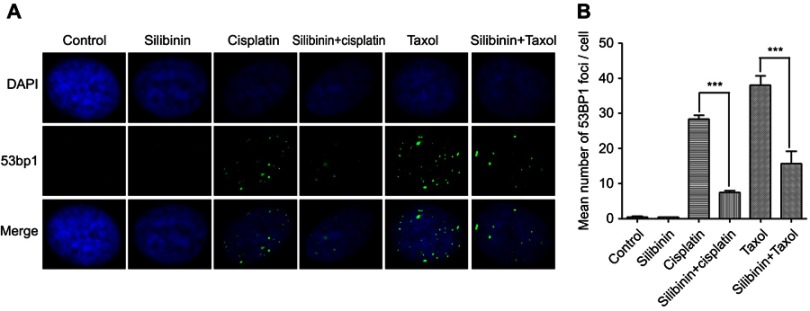
Figure 6.
Silibinin protects hepatocyte cell DNA against damage. (A) IF assay for evaluating the DNA damage. LO2 cells were treated with 0.01% DMSO (Control), silibinin (50 μM), cisplatin (24 μM), taxol (4 μM) and silibinin (50 μM) plus cisplatin (24 μM) and/or taxol (4 μM) for 48 h. DAPI and 53BP1 was the nucleus dye (blue) and DNA damage marker (green), respectively. (B) Quantification of (A). The results show the percentage of 53BP1 foci per cell among 200 untreated and treated cells, respectively. Values are the average ± SD of three independent experiments. ***p< 0.001.

