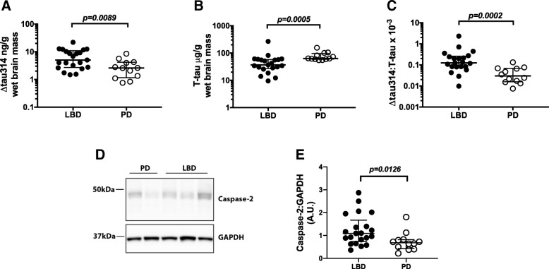
Fig. 1.
∆tau314 and caspase-2 levels are higher in LBD than PD. a-c Quantification of Δtau314 and T-tau in aqueous extracts from the superior temporal gyrus. a ∆tau314 is higher in LBD than PD. b T-tau is lower in LBD than PD. c The ∆tau314: T-tau ratio is higher in LBD than PD. d Representative western blots of Casp2 and GAPDH in aqueous extracts. e Quantification of Casp2 normalized to GAPDH. Casp2 levels are higher in LBD than PD. Data were analyzed using Mann-Whitney tests. Bars indicate the medians and interquartile ranges

