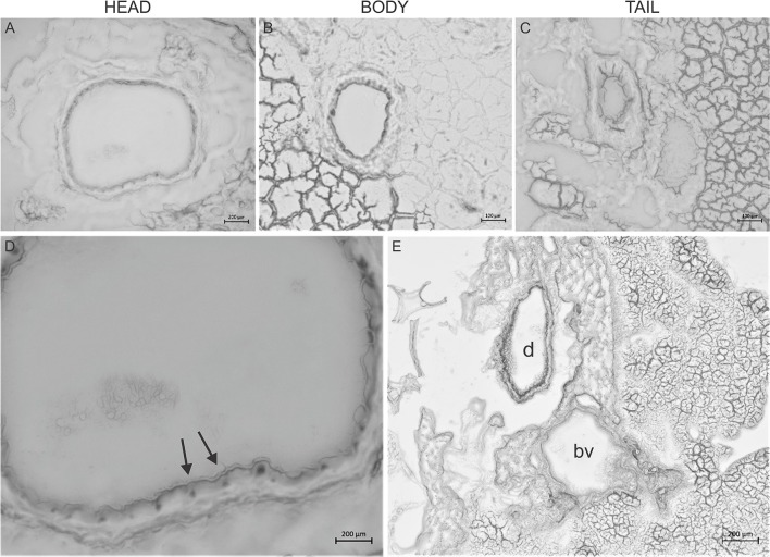
Figure 2.
Giemsa staining of the pancreas. Representative cryosections were cut from the head (A), body (B), and tail (C) of the pancreas. Giemsa stain causes dark coloring of the nuclei of inter-intralobular ducts in the head and body of the pancreas and slightly in the tail. (D) Magnified picture of (A). Arrows indicate dark coloring of the nuclei. (E) Representative cryosection from the head of the pancreas shows that Giemsa stained the duct (d) but not the blood vessels (bv).

