Figure 1.
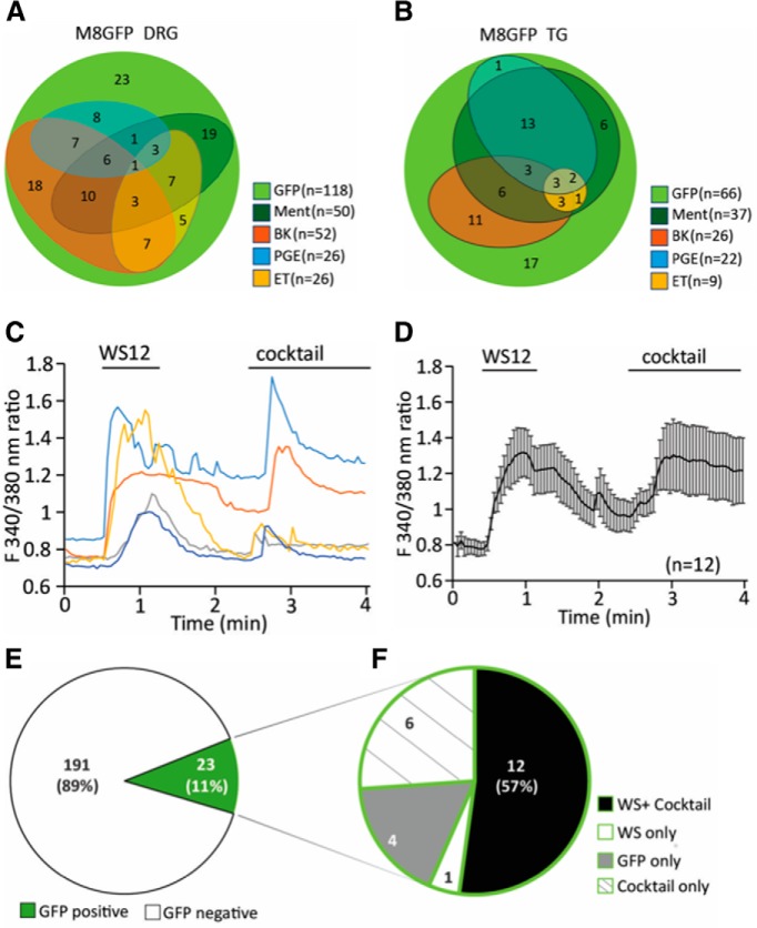
Ca2+ responses of DRG neurons to agonists of Gαq-coupled receptors and TRPM8 channels. A, Venn diagram showing the number of DRG neurons responding to 500 μm menthol, 2 μm bradykinin (BK), 1 μm PGE2, and 500 nm ET. Experiments were performed on neurons from three independent preparations from three mice both for DRG and trigeminal (TG) neurons. B, Venn diagram showing the number of TG neurons responding to the same stimuli. C, Representative traces of Ca2+-imaging measurements in TRPM8-GFP mouse DRG neurons. TRPM8 channels were activated by 10 μm WS12. A mixture containing 100 μm ADP, 100 μm UTP, 500 nm bradykinin, 100 μm histamine, 100 μm serotonin, 10 μm PGE2, and 10 μm PGI2 (cocktail) was applied to activate various Gαq-coupled receptors. Measurements were conducted in 2 mm Ca2+ NCF solution. D, Summary data of Ca2+-imaging measurements on n = 12 neurons that responded to both the cocktail and WS12 displayed as mean ± SEM. E, GFP+ neurons constitute 11% (23 neurons) of all DRG neurons (214 neurons, 2 independent preparations from 2 mice). F, Within these GFP+ neurons, 12 neurons (57%) respond to both WS12 and the inflammatory cocktail four neurons (12%) respond to neither WS12 nor cocktail six neurons (26%) only respond to cocktail and one neuron only responds to WS12.

