Figure 8.

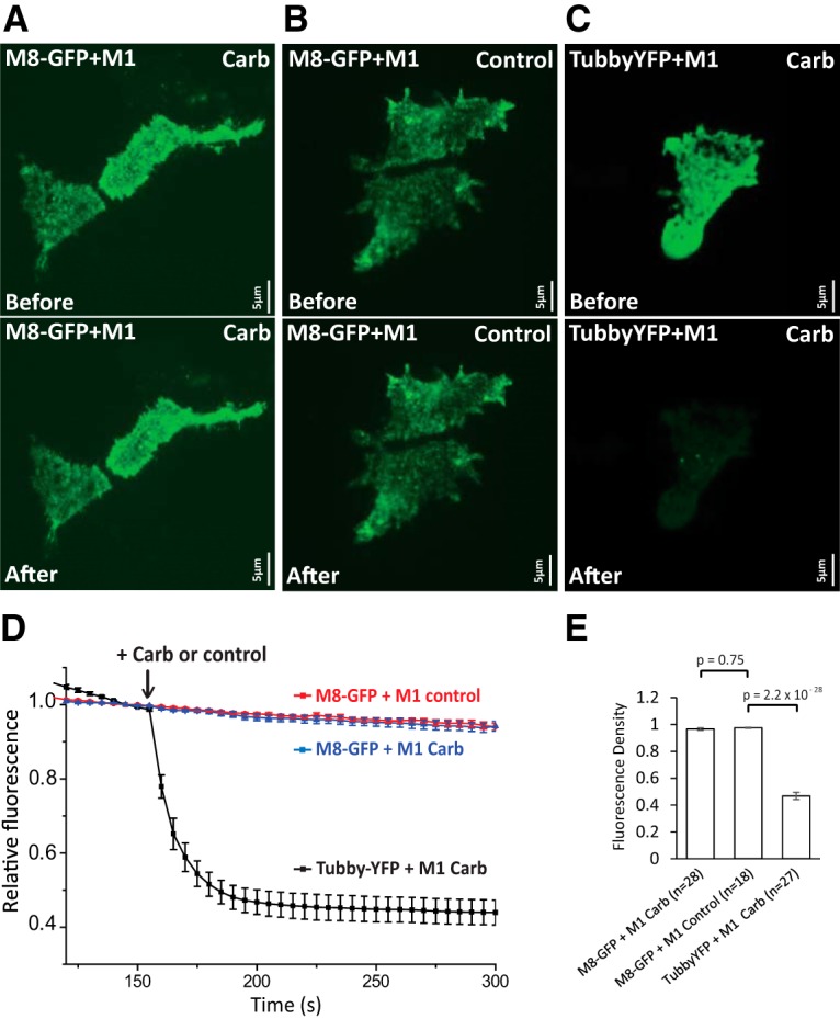
Muscarinic receptor activation does not reduce cell surface TRPM8 levels. TIRF measurements were performed as described in the Materials and Methods section in HEK293 cells transfected either with M1 muscarinic receptors and GFP-TRPM8 (M8-GFP) or M1 muscarinic receptors and the tubby YFP PI(4,5)P2 sensor. A–C are representative TIRF images before (top) and after (bottom) the application of carbachol (A, C) or no application of carbachol (B). D time course of TIRF fluorescence in the three groups, the application of 100 μm carbachol is indicated by the arrow; mean ± SEM is plotted. Fluorescence was normalized to the point before the application of carbachol. E, Summary of the three groups 50 s after the application of carbachol. Fluorescence was normalized to the point before the application of carbachol (one-way ANOVA, F(2,70) = 258.2, p = 0).
