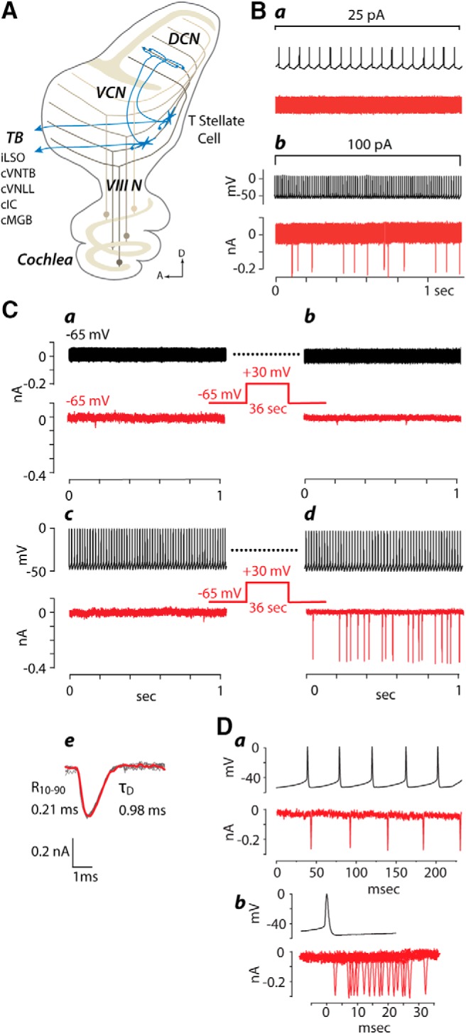
Figure 1.

A, Schematic diagram illustrating what is known about connections of T-stellate cells in the cochlear nuclei is represented in a parasagittal view. Auditory nerve fibers (VIIIN), the axons of spiral ganglion cells, carry information from the cochlea to the VCN and dorsal cochlear nucleus (DCN). The topographic pattern of innervation by these fibers imposes a tonotopic organization on the VCN and DCN; fibers tuned to low frequencies (dark brown) terminate ventrally and those tuned to high frequencies (light brown) terminate dorsally. The granule cell lamina (tan) separates the unlayered VCN from the layered DCN. The dendrites of T-stellate cells (blue stars) lie aligned with auditory nerve fibers along an isofrequency lamina. Local collateral branches of their axons terminate within the same isofrequency lamina as their dendrites in the VCN. A collateral branch innervates the same isofrequency lamina in the deep layer of the DCN. The main axon exits the VCN through the trapezoid body (TB) to innervate the ipsilateral lateral superior olive (iLSO), contralateral ventral nucleus of the trapezoid body (cVNTB), mainly contralateral inferior colliculus (cIC), and the contralateral medial geniculate body of the thalamus (cMGB). B, One of the few examples of functional interconnections between two T-stellate cells under resting conditions. Ba, A dual recording illustrates that 15 action potentials/s evoked by a small depolarizing current (25 pA) in the presynaptic cell elicited no postsynaptic currents in the second. Bb, A stronger depolarization (100 pA) that evoked 74 action potentials/s elicited EPSCs of approximately uniform size in the second. The latencies between action potentials and EPSCs were variable suggesting that EPSCs are polysynaptic. C, Pairing of presynaptic (black) firing with postsynaptic (red) depolarization potentiated an interconnection between a pair of T-stellate cells recorded simultaneously. Ca, No EPSCs were seen in either cell when the voltage of both cells was held near the resting potential. Cb, After depolarizing the postsynaptic cell to +30 mV for 36 s while the presynaptic cell continued to be clamped at −65 mV again, no responses were observed. Cc, Next, the presynaptic cell was recorded in current-clamp conditions and depolarized to fire 59 action potentials/s. Again no responses were recorded. Cd, Only when presynaptic firing was paired with depolarization were EPSCs evoked. All EPSCs in the postsynaptic cell were of approximately uniform amplitude. Dashed lines indicate that the presynaptic cell continued firing during the depolarization as before and after. Ce, 10 EPSCs are superimposed in gray and the average is superimposed in red. The 10–90% rise time (R10–90) and decay time constant (τD) of the average are shown. Da, Traces from a dual recording show that action potentials in the presynaptic cell are not locked to the timing of EPSCs in the postsynaptic cell. Db, Recordings of EPSCs aligned with respect to the action potential show that their occurrence is not locked in time. In this and in all following figures, multiple traces recorded from one cell or one pair are designated with lowercase letters. Recordings from separate cells or pairs are designated with capital letters.
